Sinus pneumatization after tooth loss reduces the vertical height of bone in the posterior maxilla. Dental implant placement for upper molar or premolar teeth may require sinus lift bone grafting to restore this lost vertical height needed to accommodate the fixture. If a large amount of bone is required, a lateral approach has been traditionally employed. This technique is invasive and postoperative recovery can be prolonged. An alternative, more minimally invasive approach can be employed, even for larger defects.
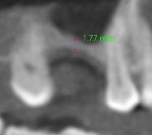
A 53 year old female presented with a severely pneumatized maxillary sinus and desire to replace tooth #13 with a dental implant. She had a lateral approach performed previously, which was abandoned due to a large sinus perforation. Pre-operative CBCT shows less than 2 mm of bone in the area of the proposed implant.
A.

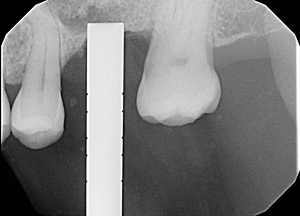
B.

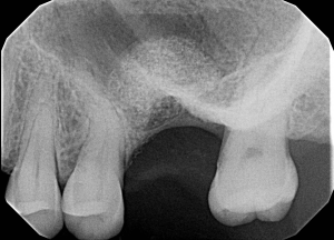
C.

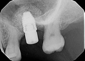
D.

Figure 1. A. Preoperative CBCT B. Sinus osteotome showing implant position C. Dome formation indicating elevation of the sinus membrane D. Final position of implant, surrounded with particulate graft.
Using a piezosurgery handpiece and sinus osteotomes, particulate graft was packed into the site. Evidence of sinus elevation was evident by the radio-opaque dome visible on an intraoperative xray. The implant was placed with good insertion torque. The patient went on to recover well with minimal pain and swelling.


